Here is a table of the large peripheral nerves that you need to know. For the practical, you should also be able to locate the four plexuses on the torso model, as well as the nerves listed below on the muscle models of upper and lower limbs (this will become easier once we go over the muscles).
The table with cranial nerves, their origin, and type of sensations perceived:
Eye-related cranial nerves:
Taste/Tongue-related cranial nerves:
Other cranial nerves:
Some cranial nerves have parasympathetic functions:
On the brain model you should be able to locate Cranial Nerve I and II, as well as name the origins of all nerves.
Showing posts with label Nervous System. Show all posts
Showing posts with label Nervous System. Show all posts
Mar 6, 2012
Feb 24, 2012
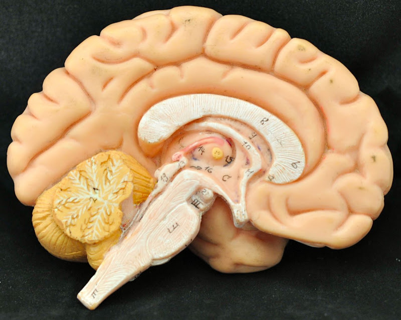
Brain Models
In the lab we have many different brain models. You should be familiar with all of them.
Start by locating the following:

On the models and in the pictures below, locate the following:
Below are two photos of the pons, medulla oblongata, and associated structures. In these pictures locate:
On the model of the fetal skull look for the sinuses:
Here is a picture of the model of the falx cerebri and associated with it straight, superior and inferior sagittal sinuses:
Below there are four pictures of the model of brain ventricles. In the pictures, try to locate the following:
Start by locating the following:
- cerebrum (cerebral hemispheres)
- frontal lobes
- parietal lobes
- temporal lobes
- occipital lobes
- cerebellum
- pons
- medulla oblongata
- convolutions (gyri, sulci, fissures)
- longitudinal fissure
- transverse fissure
- lateral sulcus
- central sulcus
- precentral gyri
- postcentral gyri

On the models and in the pictures below, locate the following:
- cerebellum
- arbor vitae
- pons
- medulla oblongata
- thalamus
- hypothalamus
- epithalamus
- optic chiasma
- pituitary gland
- pineal gland
- corpus callosum
- fornix
- septum pellucidum (visible only on most models)
- lateral ventricle (visible only on a couple of models)
- corpora quadrigemina
- superior colliculi
- inferior colliculi
- cerebral peduncles
- cerebellar peduncles
- cerebral aqueduct
- third ventricle
- fourth ventricle
- cerebral aqueduct
- choroid plexus
- falx cerebri (visible only on some models)
- superior sagittal sinus (visible only on some models)
- inferior sagittal sinus (visible only on some models)
Below are two photos of the pons, medulla oblongata, and associated structures. In these pictures locate:
- pons
- medulla oblongata
- thalamus
- optic chiasma
- corpora quadrigemina
- superior colliculi
- inferior colliculi
- cerebral peduncles
- cerebellar peduncles
- pyramids
On the model of the fetal skull look for the sinuses:
- superior sagittal sinus
- inferior sagittal sinus
- straight sinus
- transverse or horizontal sinus
- sigmoid sinus
- internal jugular vein
Here is a picture of the model of the falx cerebri and associated with it straight, superior and inferior sagittal sinuses:
Below there are four pictures of the model of brain ventricles. In the pictures, try to locate the following:
- lateral ventricles
- posterior horns
- anterior horns
- inferior horns
- third ventricle
- fourth ventricle
- lateral apertures
- median aperture
- cerebral aqueduct
- choroid plexus
Feb 23, 2012
Spinal Cord
On the model of human torso, look for following parts of the spinal cord:
On the cross section of the spinal cord look for:
- conus medullaris
- 31 pairs of spinal nerves
- cervical region of the spinal cord
- thoracic region of the spinal cord
- lumbar region of the spinal cord
- sacral region of the spinal cord
- cauda equina (horse’s tail)
- filum terminale
- dorsal root ganglion
- sympathetic ganglion
On the cross section of the spinal cord look for:
- posterior (dorsal) gray horn
- anterior (ventral) gray horn
- lateral gray horn
- gray commissure
- central canal
- dorsal root
- ventral root
- dorsal root ganglion
- dorsal rootlets
- ventral rootlets
- anterior median fissure (ventral median fissure)
- posterior median sulcus (dorsal median sulcus)
- gray matter
- white matter
Subscribe to:
Posts (Atom)


