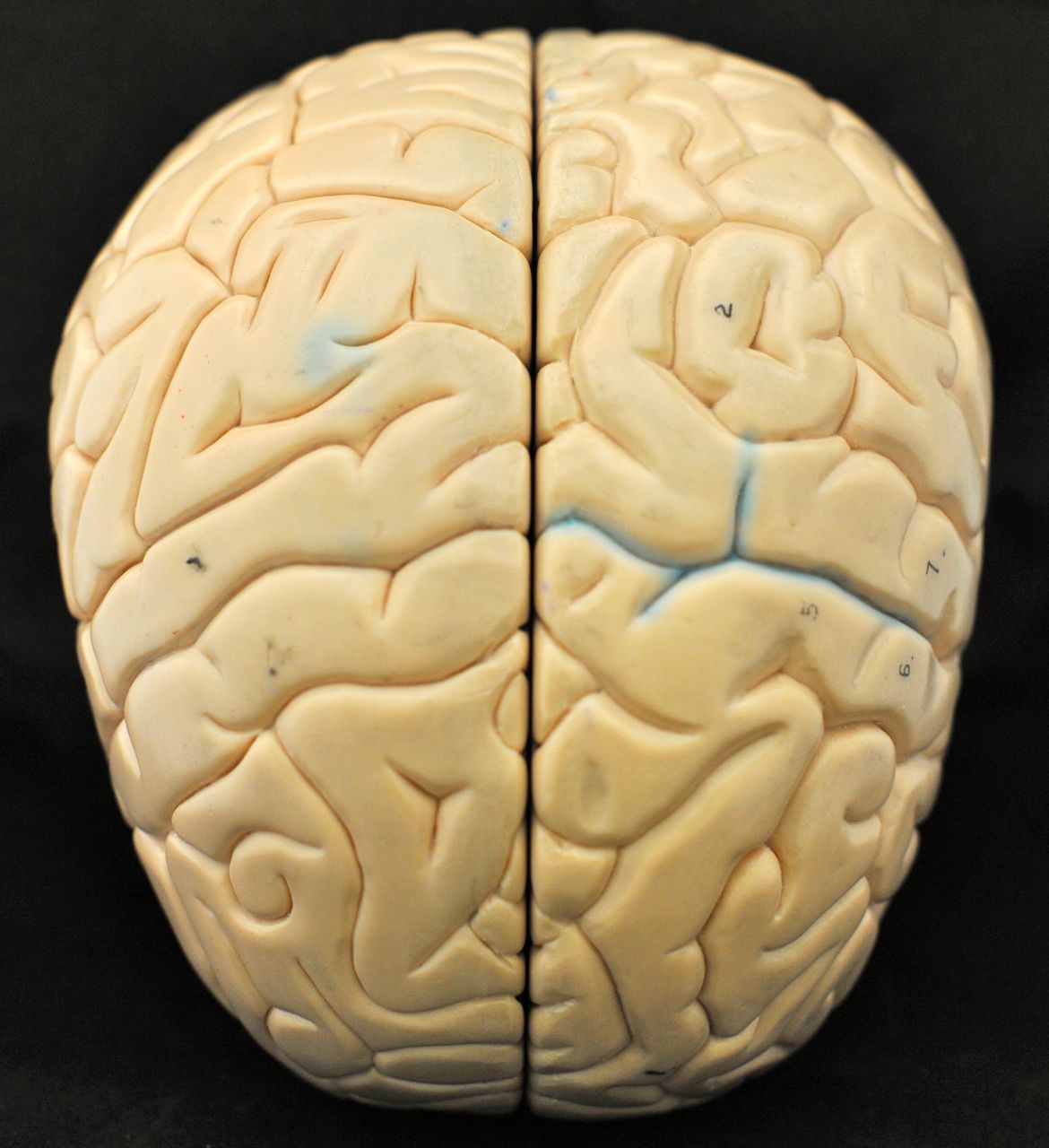
Start by locating the following:
- cerebrum (cerebral hemispheres)
- frontal lobes
- parietal lobes
- temporal lobes
- occipital lobes
- cerebellum
- pons
- medulla oblongata
- convolutions (gyri, sulci, fissures)
- longitudinal fissure
- transverse fissure
- lateral sulcus
- central sulcus
- precentral gyri
- postcentral gyri


On the models and in the pictures below, locate the following:
- cerebellum
- arbor vitae
- pons
- medulla oblongata
- thalamus
- hypothalamus
- epithalamus
- optic chiasma
- pituitary gland
- pineal gland
- corpus callosum
- fornix
- septum pellucidum (visible only on most models)
- lateral ventricle (visible only on a couple of models)
- corpora quadrigemina
- superior colliculi
- inferior colliculi
- cerebral peduncles
- cerebellar peduncles
- cerebral aqueduct
- third ventricle
- fourth ventricle
- cerebral aqueduct
- choroid plexus
- falx cerebri (visible only on some models)
- superior sagittal sinus (visible only on some models)
- inferior sagittal sinus (visible only on some models)


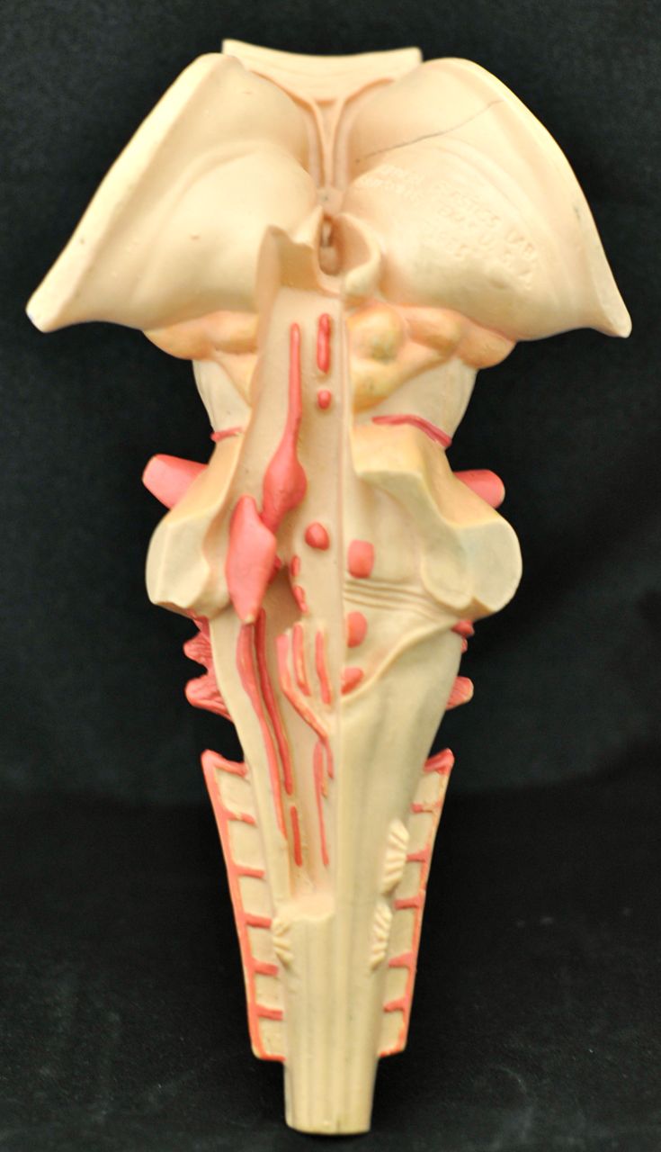
Below are two photos of the pons, medulla oblongata, and associated structures. In these pictures locate:
- pons
- medulla oblongata
- thalamus
- optic chiasma
- corpora quadrigemina
- superior colliculi
- inferior colliculi
- cerebral peduncles
- cerebellar peduncles
- pyramids

On the model of the fetal skull look for the sinuses:
- superior sagittal sinus
- inferior sagittal sinus
- straight sinus
- transverse or horizontal sinus
- sigmoid sinus
- internal jugular vein


Here is a picture of the model of the falx cerebri and associated with it straight, superior and inferior sagittal sinuses:

Below there are four pictures of the model of brain ventricles. In the pictures, try to locate the following:
- lateral ventricles
- posterior horns
- anterior horns
- inferior horns
- third ventricle
- fourth ventricle
- lateral apertures
- median aperture
- cerebral aqueduct
- choroid plexus


