Jan 31, 2012

Entrance Quiz #1 Answers

Quiz Answers for the 8 A.M. Lab. (Average score = 6.44)

  1. The heart is ventral/anterior to the spine.
  2. What is the name and the role of the marked organelle? Golgi apparatus; a sorting station: it packages, modifies, and segregates proteins for various destinations in and outside of the cell
  3. What is the name of this epithelial tissue? Where can you find it? simple cuboidal epithelium; kidney tubules (also: ovary surface, ducts of small glands)
  4. What is the name of the cavity in which the lung is located? pleural cavity
  5. What is the name and the role of the marked organelle? nucleolus; ribosome production and assembly
  6. What is the name of this epithelial tissue? Where can you find it? stratified squamous epithelium, nonkeratinized; esophagus (also: mouth, vagina)
  7. What is a chondrocyte? a cell type found in cartilage responsible for the production and maintenance of the cartilaginous matrix
  8. What is the name of this epithelial tissue? Where can you find it? simple columnar epithelium, nonciliated, with microvilla; stomach mucosa (also: the digestive tract, gallbladder)
  9. What is the name of the anatomical plane that divides the body into ventral and dorsal sections? frontal/coronal plane
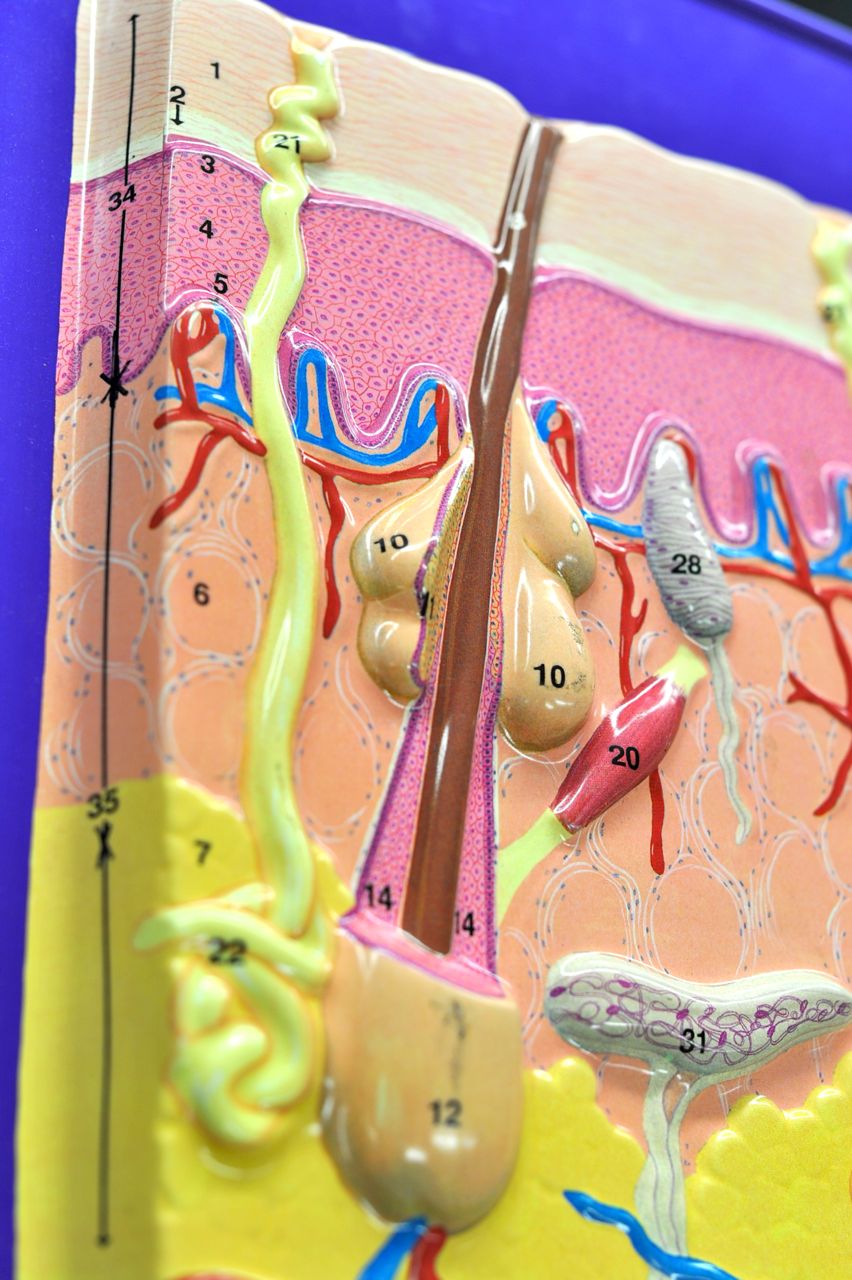
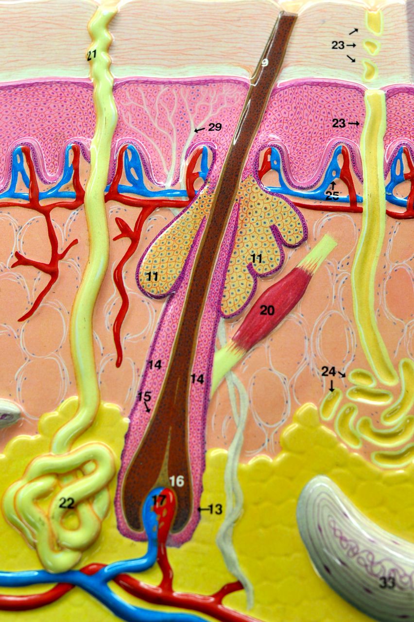
  10. What is a keratinocyte? a dominant cell type present in the epidermis  


Quiz Answers for the 10 A.M. Lab. (Average score = 7.37)

  1. The liver is ventral/anterior to the spine.
  2. What is the name and the role of the marked organelle? rough endoplasmic reticulum; proteins are synthesized on ribosomes and further modified and packaged into vesicles in rough ER
  3. What is the name of this epithelial tissue? Where can you find it? simple columnar epithelium, nonciliated, with microvilla; stomach mucosa (also: the digestive tract, gallbladder)
  4. What is the name of the membrane that lines your nose? mucous membrane
  5. What is the name and the role of the marked organelle? Golgi apparatus; a sorting station: it packages, modifies, and segregates proteins for various destinations in and outside of the cell
  6. What is the name of this epithelial tissue? Where can you find it? simple cuboidal epithelium; kidney tubules (also: ovary surface, ducts of small glands)
  7. What is a chondrocyte? a cell type found in cartilage responsible for the production and maintenance of the cartilaginous matrix
  8. What is the name of this epithelial tissue? Where can you find it? stratified squamous epithelium, nonkeratinized; esophagus (also: mouth, vagina)
  9. What is the name of the anatomical plane that divides the body into right and left sections? sagittal/median/midsagittal plane
  10. What is a keratinocyte? a predominant cell type present in the epidermis

Quiz Answers for the 2 P.M. Lab. (Average score = 6.65)

  1. My wrist is proximal to my fingers.
  2. The spleen is in the left upper abdominal quadrant.
  3. What is the name and the role of the marked organelle? smooth ER; lipid synthesis
  4. What is the name of this epithelial tissue? Where can you find it? simple cuboidal epithelium; kidney tubules (also: ovary surface, ducts of small glands)
  5. What is the name and the role of the marked organelle? mitochondrion; ATP/energy production
  6. What is this dark pink structure? nucleus
  7. What is the name of this epithelial tissue? Where can you find it? stratified squamous epithelium, nonkeratinized; esophagus (also: mouth, vagina)
  8. What is the name of this epithelial tissue? What does the arrow point to? pseudostratified columnar epithelium; cilia
  9. The cartilage cell is called the chondrocyte.
  10. Define the term: Axon An axon is a long, slender projection of a nerve cell (neuron) that typically conducts electrical impulses away from the neuron's cell body or soma.

Quiz Answers for the 4 P.M. Lab. (Average score = 6.29)

  1. My fingers are distal to my wrist.
  2. The liver is in the right upper abdominal quadrant.
  3. What is the name and the role of the marked organelle? Golgi apparatus; a sorting station: it packages, modifies, and segregates proteins for various destinations in and outside of the cell
  4. What is the name of this epithelial tissue? Where can you find it? simple cuboidal epithelium; kidney tubules (also: ovary surface, ducts of small glands)
  5. What is the name and the role of the marked organelle? nucleolus; ribosome production and assembly
  6. What is this dark pink structure? nucleus
  7. What is the name of this epithelial tissue? Where can you find it? stratified squamous epithelium, nonkeratinized; esophagus (also: mouth, vagina)
  8. What is the name of this epithelial tissue? Where can you find it? pseudostratified columnar epithelium; trachea
  9. The cartilage cell is called the chondrocyte.
  10. Define the term: Axon An axon is a long, slender projection of a nerve cell (neuron) that typically conducts electrical impulses away from the neuron's cell body or soma.

Exit Quiz #2 Answers


1.  External part of your ears is built from _____elastic cartilage____. The dominant mature cell type in this tissue is ___chondrocyte___, which lies in ___lacuna__.
2. The thick skin of palms and soles can be distinguished from the thin skin by the presence of translucent__stratum lucidum___. This skin layer lies deep to ___stratum corneum__.
3. Name the 3 types of fibers found in connective tissues: _____collagen, elastic, reticular____
4. Melanocytes can be found in stratum _____basale/germinativum_____
5. Which three (3) differences in appearance would you use to distinguish the cardiac muscle from the skeletal muscle?  ____number of nuclei; cell shape (branched vs. cylindrical); presence of intercalated discs___

6. Name the 3 types of cells found in all loose connective tissues: _____fibroblasts, fat cells, defense cells_______

Study Plan for the Entrance Quiz #2

For the next entrance quiz you need to be familiar with all the concepts we discussed today in the lab (described in the Lab Guide, Lab #2), as well as you should be able to recognize on pictures from your text the 5 items marked with (*) in the guide for the Lab #3.

Remember that it's better to study 30-60 minutes 5-6 times a week than to try to learn all the material the day before the quiz!

Here's my suggestion for how you could divide the study material between different days of the week:

Tuesday            –  The Skin + Hair + Nail
Wednesday      –  Classification of Connective Tissues; Connective Tissues Proper Loose
Thursday         –  Connective Tissues Proper Dense + Bone + Blood + Neuronal Tissue
Friday                –  Learn to recognize various Cartilages + Muscles
Saturday          –  Review what you learned on Tue and Wed
Sunday             –  Review what you learned on Thu and Fri
Monday            –  Come to the lab and ask questions at the Open Lab Review; learn the five (*) for the Lab #3

The Skin

Here's the skin model we work with in the lab. Can you identify all numbered structures on this model? How many hairs do you see on the model? How many sweat glands?


Let's zoom in on different parts of the model and try to figure out what all the numbers stand for.

First, let's look at skin layers. The skin has three principal layers:
  1. Epidermis (from Greek, "epi" means above);
  2. Dermis;
  3. Hypodermis (from Greek, "hypo" means below or low).
Can you see those layers on the model? (Clue: Look on the left side of the model.)

What does it mean that the epidermis is "nonvascular?" Can you recognize the components of the vasculature–the veins and arteries–on the model? (Side note: Arteries carry oxygenated blood to tissues, while veins drain deoxygenated blood from tissues. On all our models vessels carrying oxygenated blood are painted red, and vessels carrying oxygen-poor blood are painted blue.)


Epidermis, the outermost (or the most superficial) layer of the skin, is comprised mostly of specialized cells called keratinocytes. It can be further subdivided into four (five in the thick skin of our palms and soles) layers called strata:
  1. Stratum corneum
  2. Stratum lucidum (only present in the thick skin of our palms and soles)
  3. Stratum granulosum
  4. Stratum spinosum
  5. Stratum basale/germinativum
(Side note: strata is plural, stratum is singular.

Can you identify all those layers on the model? (Clue: Focus on numbers 1 through 5.) You should also know the key differences between those skin layers.


Dermis, the middle layer of the skin, can be divided into two parts:
  1. Papillary layer
  2. Reticular layer
On our model, there is no number associated with the papillary layer. Still, it's easy to recognize this layer, as there are many "papillae" ("pimples," from Latin), in which blood supply to the skin ends. Above this layer there is the outer nonvascular epidermis.


Let's now look at the various sensory neurons present in our skin. Can you see them on the model?

All neurons are shown in white (numbers 28 to 33). As you can see, those neurons can be present in various skin layers, and depending on the location (and type) they have slightly different functions. Some neurons are primarily involved in perception of touch, while others in perception of pain, temperature, pressure, texture, and/or stretch. You don't need to know the names of various sensory neurons, just that there are several types, and that they serve diverse functions. (If you're curious, you can read more about different sensory neurons by following these links: free nerve endings, Meissners's corpuscle, Pacinian corpuscle, Merkel nerve endings, Ruffini corpuscle.)


Now let's take a closer look at the structure of a hair. On the model below identify:
  • hair bulb
  • arrector pili muscle ("hair-raising muscle")
  • sebaceous (oil) gland associated with the hair follicle
Now look for the same three structures (hair bulb, arrector pili muscle, sebaceous gland) on the model below, and also try to locate:
  • hair root
  • hair shaft
  • hair follicle
What is the difference between hair root and hair shaft?
Hair papilla (connective tissue papilla) can only be seen in the model below. Can you see it?

In addition, the picture below shows two out of three common skin glands:
  • sebaceous gland associated with the hair follicle
  • sudoriferous (sweat) eccrine gland to the left and right of the hair follicle
The third type of skin gland is the ceruminous gland. It is not shown on the model below, because it's only present in the ear canal. There, it secretes cerumen (earwax).

(Side note: There are also apocrine sweat glands that are present mostly in the skin of the axillary and anogenital areas. Contrary to the eccrine sweat glands shown in the picture below, those glands empty into hair follicles. They are not shown on our models.)

Jan 24, 2012

Study Plan for the Entrance Quiz #1

Remember that it's better to study 30-60 minutes 5-6 times a week than to try to learn all the material the day before the quiz!

For the next entrance quiz you need to be familiar with all the concepts we discussed today in the lab (described in the Lab Guide, Lab #1), as well as you should know one-sentence definitions of the 5 items marked with (*) in the guide for the Lab #2.


Here's my suggestion for how you could divide the study material between different days of the week:

Tuesday         –   Anatomical Position, Directional Terms, Body Planes
Wednesday    –  Body Cavities and Membranes, Abdominal Quadrants
Thursday       –  Cells Components and their Functions
Friday            –  Epithelial Tissues
Saturday        –  Review what you learned on Tue and Wed
Sunday           –  Review what you learned on Thu and Fri
Monday          –  Come to the lab and ask questions at the Open Lab Review; learn the five (*) for the Lab#2

Exit Quiz #1 Answers

1. The outer serous membrane surrounding the heart is called _____parietal pericardium_________.

2. Fill in the appropriate directional term: The wrist is __distal_______ to the elbow.

3. Which cell organelle produces ATP/energy? _____mitochondrion_______.

4. The Fallopian tubes are lined with ______ (ciliated) simple columnar____ epithelial tissue. The beating of _____the cilia______ helps move the egg from the ovary to the uterus.

5. The free surface of the epithelial tissue is called the ____apical surface/membrane___, and the part that lines the bottom of the tissue is called the ______basal membrane/surface____.

Jan 23, 2012

The Cell

Below is a photo of the cell model that we use in the lab. Can you identify all structures present on this model?


What do the white dots represent?
Which organelle is shown in yellow? Which one in black?


On the model, look for:
  • endoplasmic reticulum (ER) smooth and rough - how do you distinguish between smooth and rough ER?
  • ribosomes
  • Golgi apparatus
  • nucleus - within nucleus identify nucleolus, nuclear envelope, and chromatin
  • mitochondria
  • lysosome (on our model its Yellow, as lysosome has "Y" like Yellow)
  • vacuole (on our model its Black with a gray surface)
  • microtubules/centrioles
  • cell membrane/plasma membrane/plasmalemma
Remember that you also need to know the basic function of each of these organelles.

Human Anatomy Grades

The same grade will be given for the lecture and the lab based on the cumulative points for lecture and lab.

What does it mean for you?

It means that every point counts!

Remember that you can not make up missed quizzes, so try to come to all labs.
Come on time not to miss a single entrance quiz, and stay till the very end not to miss any exit quizzes.


Typically, only 10% of students get "A" grade and 20% "B" grade. There are about 45% "Cs" and 25% "Ds" and "Fs." We want all of you to score us high as possible, and you can count on our help in achieving the grade you want. But remember that we can not study for you so, ultimately, your grade is up to you. Good luck!

Human Anatomy Lab Guidlines

1. Come On Time and Come Prepared!
  • Read the Lab Guide before the class.
>>> Familiarize yourself with new terms; mark relevant passages in your text book; identify key figures and schematics that you’ll work with during the lab session.
  • Study for the Entrance Quiz (starts at 8 or 10 A.M. sharp).
>>> Your Lab Guide will tell you what you need to know for the quiz.
>>> Typically you need to know what we just studied in the previous week + 5 items marked with (*) from the material we will study on the day of the quiz.
>>> Six BEST out of nine entrance quizzes will count towards your grade. 


2. Bring the Text and Lab Guide with you.
  • The Lab Guide will tell you what you need to know.
  • The Text will help you locate new terms on models.

3. Ask Questions if you don’t understand the material, or if you are not sure if you identified all the terms correctly. 
 

4. At home, study several times a week, preferably with a classmate.

>>> Quiz and challenge each other. 
>>> Write down any questions you might have and ask them during the next class or Monday Night Open Lab Review.

>>> Remember that memory works just like muscles: You cannot win a marathon if you only start preparing for it a day before. You need to exercise your muscles (and memory) by "working out" several times a week, at regular intervals. Our brains also work best after a good night's sleep and a hearty breakfast. So instead of burning the midnight oil just before your class/exam, better start studying a few days in advance.  


5. Come to Open Lab Review sessions on Monday evenings (7:00 P.M. to 9:00 P.M.).