Feb 28, 2012

Exit Quiz #5 Answers


1.     CSF is found between which two layers of the meninges? Arachnoid and pia maters.
2.     The floor of the 3rd ventricle is formed mostly by the __hypothalamus.
3.     Arbor vitae refers to the appearance of the __ white matter/myelinated axons ___ in the __cerebellum.
4.     Each of the ventricles has an associated structure called a ___ choroid plexus_, which produces __ CSF ___.
5.     The ___ corpora quadrigemina____ coordinates head movement with visual and auditory stimuli.

Study Plan for the Entrance Quiz #4

For the next entrance quiz you need to be familiar with all the concepts we discussed today in the lab (described in the Lab Guide, Lab #5 part 1), as well as you should know definitions and be able to recognize on pictures from your text the 5 items marked with (*) in the guide for the Lab #5 part 2 and Lab #6.


Here's my suggestion for how you could divide the study material between different days of the week:

Tuesday            –   Subdivisions of the Nervous System and General Characteristics of the CNS
Wednesday      –  Spinal Cord
Thursday         –  Forebrain, Midbrain and Hindbrain
Friday               –  Ventricles of the Brain + Brain Meninges
Saturday          –  Review what you learned on Tue and Wed
Sunday             –  Review what you learned on Thu and Fri
Monday            –  Come to the lab and ask questions at the Open Lab Review; learn the five (*)

Remember that it's better to study 30-60 minutes 5-6 times a week than to try to learn all the material the day before the quiz!

Feb 24, 2012

Brain Models

In the lab we have many different brain models. You should be familiar with all of them.

Start by locating the following:
  1. cerebrum (cerebral hemispheres)
    • frontal lobes
    • parietal lobes
    • temporal lobes 
    • occipital lobes
  2. cerebellum
  3. pons
  4. medulla oblongata
  5. convolutions (gyri, sulci, fissures)
    • longitudinal fissure
    • transverse fissure
    • lateral sulcus
    • central sulcus
    • precentral gyri
    • postcentral gyri


On the models and in the pictures below, locate the following:

  • cerebellum
  • arbor vitae
  • pons
  • medulla oblongata
  • thalamus
  • hypothalamus
  • epithalamus
  • optic chiasma
  • pituitary gland
  • pineal gland
  • corpus callosum
  • fornix
  • septum pellucidum (visible only on most models)
  • lateral ventricle (visible only on a couple of models)
  • corpora quadrigemina
  • superior colliculi
  • inferior colliculi
  • cerebral peduncles
  • cerebellar peduncles
  • cerebral aqueduct
  • third ventricle
  • fourth ventricle
  • cerebral aqueduct
  • choroid plexus
  • falx cerebri (visible only on some models)
  • superior sagittal sinus (visible only on some models)
  • inferior sagittal sinus (visible only on some models)


Below are two photos of the pons, medulla oblongata, and associated structures. In these pictures locate:
  • pons
  • medulla oblongata
  • thalamus
  • optic chiasma
  • corpora quadrigemina
  • superior colliculi
  • inferior colliculi
  • cerebral peduncles
  • cerebellar peduncles
  • pyramids


On the model of the fetal skull look for the sinuses:
  • superior sagittal sinus
  • inferior sagittal sinus
  • straight sinus
  • transverse or horizontal sinus
  • sigmoid sinus
  • internal jugular vein


Here is a picture of the model of the falx cerebri and associated with it straight, superior and inferior sagittal sinuses:

Below there are four pictures of the model of brain ventricles. In the pictures, try to locate the following:
  1. lateral ventricles
    • posterior horns
    • anterior horns
    • inferior horns
  2. third ventricle
  3. fourth ventricle
    • lateral apertures
    • median aperture
  4. cerebral aqueduct
  5. choroid plexus


Feb 23, 2012

Spinal Cord

On the model of human torso, look for following parts of the spinal cord:
  • conus medullaris
  • 31 pairs of spinal nerves
  • cervical region of the spinal cord
  • thoracic region of the spinal cord
  • lumbar region of the spinal cord
  • sacral region of the spinal cord
  • cauda equina (horse’s tail)
  • filum terminale
  • dorsal root ganglion
  • sympathetic ganglion


On the cross section of the spinal cord look for:

  • posterior (dorsal) gray horn
  • anterior (ventral) gray horn
  • lateral gray horn
  • gray commissure
  • central canal
  • dorsal root
  • ventral root
  • dorsal root ganglion
  • dorsal rootlets
  • ventral rootlets
  • anterior median fissure (ventral median fissure)
  • posterior median sulcus (dorsal median sulcus)
  • gray matter
  • white matter


Feb 21, 2012

Practical #1 Questions and Answers

The average score for the lab section #1 (8 A.M.) was only 54%, and for the section #2 (10 A.M.) 63%. These are the lowest scores we've ever seen on any practical . . . You should get your individual scores by the end of the week, after all lab instructors grade their groups, and we adjust the scores.

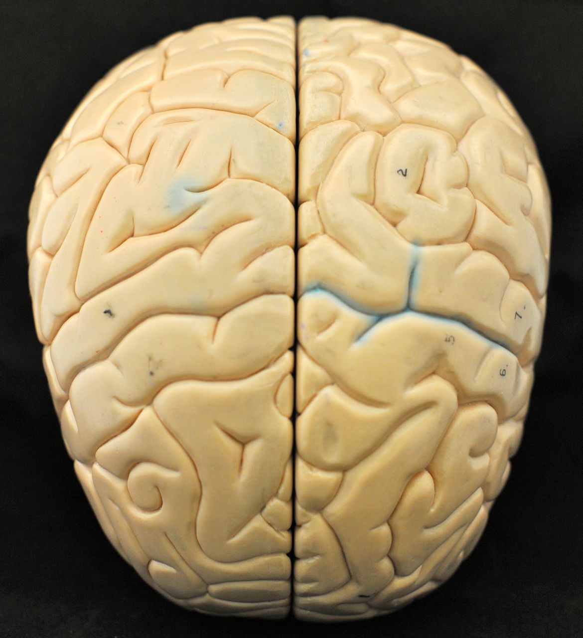
In meantime, I would like to encourage you to start studying for the next lab. (We'll start working on the brain! We might even get some real human brains for you.) There won't be an entrance quiz at the beginning of the lab, but it's always a good idea to come prepared for the class. That way you'll use your time in the lab more efficiently. At the very minimum familiarize yourself with the following figures from the Chapter 12 in your text: 12.6; 12.12; 12.15; and 12.31.


Practical #1 Questions and Answers


STATION 1 (Scapula)
1.     Identify A – Coracoid process
2.     Identify B – Supraspinous fossa
3.     Identify C – Medial border

STATION 2 (Atlas + Axis)
1.     What does A articulate with? – Occipital condyles
2.     Identify B – Dens
3.     What movement occurs at the joint between C1 and C2? – Head rotation/shaking (Not nodding)

STATION 3 (Humerus)
1.     Identify A – Deltoid tuberosity
2.     Identify B – Capitulum
3.     Identify C – Coronoid fossa

STATION 4 (Torso Model)
1.     Name the cavity in which A is located? – Pleural cavity
2.     What kind of membrane lines A? – Serous membrane
3.     What plane is this? – Midsagittal/Sagittal plane

STATION 5 (Midsagittal section of the skull)
1.     Identify A – Frontal sinus
2.     Identify B – Internal acoustic/auditory meatus
3.     In which bone is B located? – Temporal bone (petrous portion of the temporal bone)

STATION 6 (Hip Bone)
1.     Identify A – Acetabulum
2.     Identify B – Greater sciatic notch
3.     Identify C – Auricular surface

STATION 7 (Tissue: Skin)
1.     Identify layer A – Stratum granulosum
2.     Identify layer B – Stratum spinosum
3.     Which pigment-containing cells can you find in the layer C? – Melanocytes

STATION 8 (Roof of the cranium, Fetal skull)
1.     Identify A – Lambdoidal/lambdoid suture
2.     Identify B – Fontanelle
3.     Identify C – Occipital bone (Basioccipital bone)

STATION 9 (Femur, Patella)
1.     Identify A – Lesser trochanter
2.     Identify B – Lateral epicondyle
3.     Identify C – Apex of patella

STATION 10 (Vertebrae)
1.     What kind of tissue is A made of? – Fibrocartilage
2.     What is the primary cell type found in this tissue? – Chondrocyte/Chondroblast
3.     What is the primary fiber type found in this tissue? – Collagen

STATION 11 (Skin model)
1.     Identify A – Sweat/Sudoriferous gland
2.     Identify B – Arrector pili muscle
3.     Identify C – Nerve/Nerve ending/Pacinian corpuscule

STATION 12 (Ulna)
1.     Identify A – Radial notch (of ulna)
2.     Identify B – Olecranon (process)
3.     Is it left or right bone? Left

STATION 13 (Tissue: Bone)
1.     What goes through A? – Blood vessels and nerves (both answers needed)
2.     Identify B – Lamellae
3.     What is the primary fiber type found in this tissue? – Collagen

STATION 14 (Ribs, Lumbar vertebra)
1.     Identify A – Xiphoid process
2.     What kind of tissue is B made of? – Hyaline cartilage
3.     Identify C – Spinous process of the lumbar vertebra (both parts of the answer needed)

STATION 15 (Pelvis)
1.     Identify A – Pubic symphysis
2.     Identify B – Anterior inferior iliac spine
3.     Identify C – Ischial ramus

STATION 16 (Mandible, Skull)
1.     Identify A – Symphysis of the mandible/Mandibular symphysis/Symphysis menti
2.     Identify B – Petrous portion of the temporal bone (both parts of the answer needed)
3.     Identify C – Maxilla

STATION 17 (Tissues: Simple columnar w/microvilla #1 and Psuedostratified columnar w/cilia #2)
1.     Which of these two tissues has cilia? – #2
2.     Where can you find the tissue #2? – Trachea, or Upper Respiratory Tract/Nasal Cavity/Larynx/Pharynx (Sperm ducts/Vas deferens/Ductus deferens have non-ciliated pseudostratified ephitelium; Epidydymis have stereocilia)
3.     Identify C – Goblet cells

STATION 18 (Tibia, Fibula)
1.     Identify A – Intercondylar eminence
2.     Identify B – Medial malleolus of tibia
3.     Identify C – Head of fibula (both parts of the answer needed)

STATION 19 (Rib, Cervical vertebra)
1.     Identify A – Transverse foramen of the cervical vertebra (both parts of the answer needed)
2.     Identify B – Head of the rib (both parts of the answer needed)
3.     What does B articulate with? – Coastal facets/Coastal demi-facets/ Superior and inferior coastal demi-facets/body of the thoracic vertebra (both parts of the answer needed)

STATION 20 (Cell Model)
1.     Identify A – Mitochondria
2.     Identify B – Golgi
3.     What cellular process is C involved in? – Mitosis/Cell division/Microtubule organization/Cytokinesis

STATION 21 (Foot)
1.     Identify A – Navicular
2.     Identify B – #4 (right) metatarsal
3.     How many phalanges are in the right foot? – 14

STATION 22 (Skull)
1.     Identify A – Superior nuchal line
2.     Identify B (the indentions) – Cribiform plate (Not Crista galli)
3.     What does pass through the openings in B? – Olfactory nerves

STATION 23 (Skull)
1.     Identify A – Nasal bone
2.     Identify B – Greater wing of the sphenoid bone (both parts of the answer needed)
3.     Part of which bone is C? – Temporal bone (Not zygomatic bone)

STATION 24 (Radius, Clavicle)
1.     Identify A – Styloid process of radius (both parts of the answer needed)
2.     Identify B – Radial tuberosity (of radius)
3.     Identify C – Sternal end of the clavicle (both parts of the answer needed)

STATION 25 (Tissue: Skin)
1.     Identify tissue type (A) – Keratinized stratified squamous epithelium (full description needed)
2.     Identify tissue type (B) – Dense irregular/Connective tissue
3.     What is the primary function of the tissue A? – Protection/Moisture maintenance

Feb 15, 2012

The Skull

Thanks to Thi for sharing those great photos of the skull!

Open Lab times on the weekend:
Saturday 10 A.M. to 12 P.M. (Thi)
Sunday 1 P.M. to 7 P.M. (1 P.M. to 4 P.M. Laurie, 4 P.M. to 7 P.M. Monika)


On the model below, look for:
  • Crista galli and cribriform plate of the ethmoid bone
  • Greater and lesser wings of the sphenoid bone
  • Sella turica of the sphenoid bone
  • Optic canals
  • Jugular foaraminas
  • Foramen magnum
  • Hypoglossal canals
  • Internal acoustic meatus

On the model below, look for:
  • Crista galli and cribriform plate of the ethmoid bone
  • Greater and lesser wings of the sphenoid bone
  • Sella turica of the sphenoid bone
  • Optic canals
  • Jugular foaramina
  • Foramen magnum
  • Hypoglossal canals
  • Internal acoustic meatus  
  • Petrous portion of the temporal bone


On the model below, identify:
  • Crista galli and cribriform plate of the ethmoid bone
  • Greater and lesser wings of the sphenoid bone
  • Sella turica of the sphenoid bone
  • Optic canals
  • Jugular foaraminas
  • Foramen magnum
  • Hypoglossal canals
  • Internal acoustic meatus


On the model below, identify:
  • Crista galli and cribriform plate of the ethmoid bone
  • Greater and lesser wings of the sphenoid bone
  • Sella turica of the sphenoid bone
  • Optic canals

On the model below, identify:
  • Squamous suture
  • Squamous region of the temporal bone
  • Tympanic region of the temporal bone
  • Mastoid region of the temporal bone
  • Mastoid process 
  • External acoustic meatus
  • Styloid process
  • Zygomatic process of the temporal bone
  • Zygomatic bone
  • Mandibular fossa
  • Maxilla
  • Alveolar margin of the maxilla
  • Frontal bone
  • Occipital bone
  • Parietal bone
  • Pterygoid process of the sphenoid bone


On the model below, identify:
  • Maxillary bones
  • Alveolar margin of the maxilla
  • Frontal bone
  • Zygomatic bones
  • Vomer
  • Perpendicular plate of the ethmoid bone
  • Inferior nasal conchae
  • Middle nasal conchae (of the ethmoid bone)


On the model below, identify:

  • Maxilla
  • Palatine bones
  • Vomer 
  • Pterygoid process of the sphenoid bone
  • Mandibular fossa
  • Mastoid process 
  • External acoustic meatus
  • Styloid process
  • Zygomatic process of the temporal bone
  • Zygomatic bone
  • Parietal bones
  • Occipital bone
  • Occipital condyles
  • Occipital protuberance
  • Foramen magnum
  • Jugular foaramina

On the model below, identify sutures and bones they connect.


On the model below, examine the parts of ethmoid bone and its location in the relation to other bones in the skull.


On the model below, examine the structure of the maxilla and palatine bones.


On the model below, examine the parts of ethmoid and sphenoid bones and their location in the relation to each other and other bones of the skull.


On the model below, look for the frontal, sphenoid and maxillary sinuses. Also, identify vomer.


The next two models show the neonatal skulls. Can you remember the major differences between the adult and neonatal skulls?


Feb 14, 2012

Exit Quiz #4 Answers


1.  Posteriorly, all ribs attach to the thoracic vertebrae. The head of the rib articulates with _______the body of the same-numbered and immediately superior vertebrae___________________, and the tubercle of the rib articulates with  _____the costal facet of the transverse process of the same-numbered vertebra _______
2. In the anatomical position which of the forearm bones lies medially? __ulna___________________

3. How many phalanges are there in the right thumb? ___2___How is each specifically identified in anatomical terms? ________right #1 proximal phalanx; right #1 distal phalanx___________________
4. Proximally, the femur articulates with ________the hip bone____________________________, and distally, it articulates with ______the tibia________
5. Compare the model of the female and the male pelvis and identify the anatomical childbearing adaptations of the ischial tuberosities, sacrum and coccyx. ____________ischial tuberosities shorter; sacrum wider and shorter; coccyx straighter, movable______________________________________

Extra Question, not graded.
6. Which bones contribute to the knee joint?  _______femur, tibia, and patella______________ 

Study Plan for the Practical #1

The Practical #1 is only one week away! If you haven't started studying for it yet, it's the highest time that you do.

There will be 75 questions on the practical, each worth one point. These 75 questions will be divided between 25 "stations." Each station will have a model (e.g. a skull, a cell model, a bone, e.t.c.) and there will be three questions associated with the featured model. The majority of questions will be "Identify A," "Identify B," "Identify C,"–where "A," "B," and "C" will be marked on the model. But occasionally there might be other questions too, for example: (1) We might ask a type of cell/fiber that can be found in different tissues or we can ask where in the body the tissue can be found; (2) We can ask about a function of some of the cellular organelles; (3) We can ask with which other bone the bone in question articulates (connects), or if it's left or right bone. Still, the majority of questions will simply ask you to identify what you can see, so you should first study these kinds of questions.

Typically, on the practical there are more questions from the last lab, as this is the one from which you were not quizzed yet on the entrance quiz. So on the upcoming practical you should expect a lot of questions about appendicular skeleton. There will be a lot of questions about the skull, too. Study at home as much as you can, and please come on Saturday and Sunday to the Open Lab Review to look at the models. It's impossible to learn all those difficult concepts just from the book, without carefully examining the models. I'll be in the lab on Sunday afternoon (4 P.M. to 6 P.M.), so please come and ask me as many questions as you want! (Likely, on Sunday the lab will be open 4 P.M. to 8 P.M., and it will also be open on Saturday afternoon, probably after 5 P.M. I will update the times once I know them. They will be on the blackboard too.) 

Good luck!  


Study Plan for the Practical #1

TODAY (during and after the class):               Appendicular skeleton (lab 4)
WEDNESDAY:                                                          Appendicular skeleton (lab 4)
THURSDAY:                                                              Axial skeleton (lab 3)
FRIDAY:                                                                    Anatomical terms, Cells, Tissues + Skin (lab 1+2)
SATURDAY:                                                             Open Lab – Review Appendicular skeleton (lab 4)
SUNDAY:                                                                 Open Lab - Review Axial skeleton (lab 3)
MONDAY:                                                               No Open Lab. Review Labs 1+2+3+4

1) Study at least 3-4 hours a day.
2) While you study, make a list of questions. Ask those questions to other students and the lab instructor during the Open Lab Review.
3) I'll be in the lab on Sunday afternoon 4 P.M. to 6 P.M. You can get back your entrance and exit quizzes then.

Entrance Quiz #3 Answers

Quiz Answers for the 8 A.M. Lab:

  1. What is a suture? It is a type of non-movable fibrous joint (synarthrosis) made of a thin layer of dense fibrous connective tissue that unites skull bones. What bones are joined by the squamous suture? the parietal and temporal bones 
  2. Which sinus is shown on the picture below? the sphenoid
  3. What is the name of this bone? the temporal bone Name A and B. A: zygomatic process B: external acoustic meatus
  4. What is the name of this bone? mandible Name A and B. A: alveolar margin B: mental foramen
  5. What is the name of this bone? the zygomatic bone
  6. Identify A. crista galli
  7. Identify A. jugular foramen
  8. Identify A and B. A: optic canal B: inferior orbital fissure
  9. What is the name of this bone? (lumbar) vertebrae What is A? (superior) articular process
  10. What is the name of this bone? humerus What is A? trochlea

Quiz Answers for the 10 A.M. Lab:

  1. What is a suture? It is a type of non-movable fibrous joint (synarthrosis) made of a thin layer of dense fibrous connective tissue that unites skull bones. What bones are joined by the lambdoid suture? the parietal, temporal, and occipital bones
  2. Which sinus is shown on the picture below? the frontal
  3. What is the name of this bone? the temporal bone Name A and B. A: squamous region B: external acoustic meatus
  4. What is the name of this bone? mandible Name A and B. A: alveolar margin B: mental foramen
  5. What is the name of this bone? maxilla
  6. Identify A. crista galli
  7. Identify A. sella turica
  8. Identify A and B. A: optic canal B: superior orbital fissure
  9. What is the name of this bone? (lumbar) vertebrae What is A? (superior) articular process
  10. What is the name of this bone? humerus What is A? trochlea

Feb 8, 2012

Exit Quiz #3 Answers


1.  What bones are joined by the squamous suture? _____temporal and parietal_________________
2. The nasal septum is composed of a single cartilage component ___the septal cartilage__________, and two major bony components __vomer and perpendicular plate of ethmoid bone_________________ (be specific: X part of Y bone).

3. The atlas articulates with__the occipital condyles (of the occipital bone)__ (be specific)
4. The human skull has numerous foramina (holes) through which nerves, arteries and veins pass.  For example, the hypoglossal canals located in the ________occipital bone___________ transmit ______hypoglossal nerve________, which controls almost all muscles of your tongue, thereby controlling speech, food manipulation, and swallowing.
5. Name four pairs of paranasal sinuses:  ____frontal, sphenoid, ethmoid, maxillary_______________

Feb 7, 2012

Study Plan for the Entrance Quiz #3 and Practical #1

Yes, it's time to start studying for the practical. It's only two weeks away . . . 

For the next entrance quiz you need to be familiar with all the concepts we discussed today in the lab (described in the Lab Guide, Lab #3, part 1), as well as you should be able to recognize on pictures from your text the 5 items marked with (*) in the guide for the Lab #3 part 2 and Lab #4.

From now on till the practical you should be studying at least two hours every day.

Here's my suggestion for how you could divide the study material between different days of the week:

Tuesday            –  Names and general location of all skull bones + Sutures;
Wednesday      –  Identify parts and region of bones on the pictures of individual bones;
Thursday         –  Identify parts and region of bones on the various sections of the skull;
Friday               –  Vertebral column + Parts of vertebrae + Differences between different vertebrae + Rib Cage
Saturday          –  Review what you learned on Tue, Wed, Th and Fri
Sunday             –  A quick review of Labs #1 and #2 (Tissues + Skin)
Monday            –  Come to the Open Lab Review and work with the skull models we have in the lab + Learn the five (*) for the Lab #3 part 2 and Lab #4

Entrance Quiz #2 Answers

  1. What is the name of this tissue? Which two primary types of cells can you find in this tissue? connective tissue proper: loose, adipose tissue; fat cells (adipose cells) and fibroblasts
  2. Crista galli is part of which bone? the ethmoid bone
  3. Name A-C on the pictures: A axon B dendrite C neuroglia/supporting cells/Schwann cells
  4. What is the name of this tissue? Where can you find it? connective tissue proper, dense irregular; fibrous capsules of organs and joints, dermis of the skin, submucosa of digestive tract
  5. List layers of epidermis of THIN skin starting with the most superficial one. stratum corneum, stratum granulosum, stratum spinosum, stratum basale
  6. What is the name of this tissue? What is A? bone/osseous tissue; Haversian/central canal
  7. What is the name of this tissue? cardiac muscle
  8. Name A-B on the picture: A arrector pili muscle B sebaceous/oil gland
  9. What is the name of this tissue? What is A? elastic cartilage; elastic fibers (matrix also ok)
  10. What is A? foramen/foramina

Feb 1, 2012

Tissues

Animal tissues can be grouped into four basic types:
  1. epithelial
  2. connective
  3. muscle
  4. nervous


The epithelial tissues provide a barrier between the external environment and our organism. You can find this tissue on the surface of all organs that are in contact with the outside environment, such as: the skin, the airways, the inner lining of the digestive tract, and the reproductive tract.

Epithelial tissue helps to protect our organism from microorganisms, injury, and fluid loss. In addition to this protective function, epithelial tissue may also be specialized to function in secretion and absorption.


As the name implies, connective tissue serves a "connecting" function. It supports and binds other tissues, it gives shape to our organs and holds them in place. Examples of this tissue include: bone, cartilage, tendons, ligaments, adipose tissue, blood, lymphatic tissue, and others.

Connective tissue has three main components: cells, fibers, and extracellular matrix.


Muscle tissue is the active contractile tissue of our body, which produces force and causes motion.

There are three distinct classes of muscle tissue:
  • smooth muscle, which is found in the inner linings of organs;
  • skeletal muscle, which is found attached to bones and enables us to move;
  • cardiac muscle, which is found in the heart, allowing it to pump blood throughout our body.


Nervous tissue is the main component of the central (the brain and spinal cord) and peripheral nervous systems (the cranial and spinal nerves). The nervous system functions include sensory input, integration, control of muscles and glands, homeostasis, and mental activity.

Nervous tissue is composed of neurons, which transmit impulses, and the neuroglia (supporting cells), which assist in propagation of the nerve impulse and provide nutrients to the neurons.


Last week you learned how to recognize different types of epithelial tissues. Your task for this week is to learn how to recognize different types of muscles and connective tissues. You should work with the pictures in your text and the blackboard (you can download from there slides of all the tissues you saw in the lab). You can also come to the open lab on Monday evening and look at all these slides on our projectors. These slides will be used for the practical exam.


I'm going to give you a few "quick and dirty" tips on how to recognize different types of connective tissue, and how to determine what kind of cells and fibers are present there.

Connective tissues can be classified into four main types: blood, bone, cartilage, and connective tissue proper. I'm sure you have no problem recognizing "blood" and "bone," so I'm going to focus on cartilage and connective tissue proper. Just remember that the key cell type present in bone is osteocyte/osteoblast, whereas in blood you can find erythrocyte, white blood cells, and thrombocytes.

  1. After you decided that what you see is not blood, bone, muscle, nervous, or epithelial tissue, now you have to determine if it is cartilage or connective tissue proper. One way of telling them apart is by the cell shape: since chondrocytes (the cells present in the cartilage tissue) lie in spaces called lacunae, they usually have trapezoidal or semi-circular shape. If you see cells like this, likely you're dealing with cartilage.
  2. There are three types of cartilage tissue: hyaline, elastic, and fibro-cartilage. You can quite easily tell them apart by looking at the matrix: (1) the hyaline cartilage matrix is glassy in appearence and there are no visible fibers; (2) the elastic cartilage has dark thin elastic fibers around the lacunae; and (3) the fibro-cartilage is named like this because there are lots of thick purple collagen fibers in the matrix.
  3. Remember, in all cartilage tissues the dominant cell type is chondrocyte (which lies in lacuna).
  4. As the name suggests, the dominant fiber type in the elastic cartilage is the elastic fiber, whereas in the two other types of cartilage it's collagen.
  5. If the cell shape is not trapezoidal or semicircular, likely you're looking at the connective tissue proper. The first step is to decide if it's connective tissue proper "dense" or "loose." If you look carefully at the histological sections you'll see that the "loose" tissues have some empty, white spaces between cells, whereas in the "dense" tissues cells are a bit squished and their nuclei are also squished and look more like discs than circles.
  6. Remember, in all connective tissues proper, except for the adipose tissue, the dominant cell type is fibroblast. There are fibroblasts in the adipose tissue too, but they're in the minority compared to fat cells.
  7. If you see the squished cells and nuclei, you're looking at the connective tissue proper dense. Now you have to decide if it's "regular," "irregular," or "elastic."
  8. The connective tissue proper dense elastic has characteristic dark elastic fibers, whereas the connective tissues proper regular and irregular have collagen fibers, which are lighter in color (usually pinkish) and thicker. If those fibers are nicely organized and all align in one direction, it's connective tissue proper regular. If the fibers look messy, it's connective tissue proper irregular.
  9. Again, remember the dominant fiber type in the connective tissue proper elastic is elastic (it makes sense, doesn't it?), whereas in the two other types of connective tissues proper it's collagen. The dominant cell type in all those tissue is fibroblast.
  10. Now, there are three types of connective tissue proper loose. I think you can recognize the adipose tissue quite easily? Remember that those round cells are adipocytes/fat cells and they're filled with fat. In fact there is so much fat there, that all other organelles are pushed to the site. Over 90% of this tissue is fat cells, but there are some fibroblasts there too. In the end, it's the connective tissue proper.
  11. The last two tissues that we need to learn to distinguish from each other are the connective tissues proper loose "reticular" and "areolar." The connective tissue proper loose reticular will have many dark reticular fibers (which often align to form a mesh/net), whereas in the connective tissue proper loose areolar you can see all three types of fibers: the thick purple collagen, the thinner elastic fiber, and the thinnest, darkest reticular fiber.
  12. In summary, remember that in all connective tissues proper you can find fibroblasts, and in all cartilage tissues you can find chondrocytes. In most of these tissues the dominant fiber type is collagen (except for the ones that have "elastic" or "reticular" in their names). So if you're asked about the fiber type and you're not sure what's the right answer, the safe bet is to say "collagen." (Again, unless the tissue name suggests that it has "elastic" or "reticular" fibers. Also, areolar tissue has all three types of fibers clearly visible.)
  13. Hope that helps! Don't forget also to learn where each of these tissues is located. The best strategy is to think how the tissue's properties (cell type, type and arrangement of fibers) influence its function/location in the body.


Below is the summary of slides I showed you yesterday in the lab. You can download and print them if you want.