Here's the skin model we work with in the lab. Can you identify all numbered structures on this model? How many hairs do you see on the model? How many sweat glands?

Let's zoom in on different parts of the model and try to figure out what all the numbers stand for.
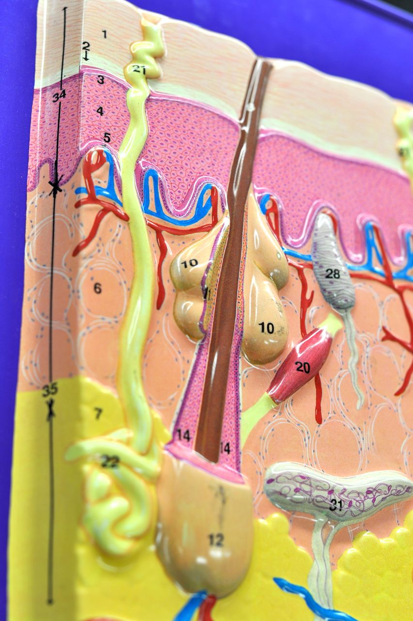
First, let's look at skin layers. The skin has three principal layers:
What does it mean that the epidermis is "nonvascular?" Can you recognize the components of the vasculature–the veins and arteries–on the model? (Side note: Arteries carry oxygenated blood to tissues, while veins drain deoxygenated blood from tissues. On all our models vessels carrying oxygenated blood are painted red, and vessels carrying oxygen-poor blood are painted blue.)
Epidermis, the outermost (or the most superficial) layer of the skin, is comprised mostly of specialized cells called keratinocytes. It can be further subdivided into four (five in the thick skin of our palms and soles) layers called strata:
Can you identify all those layers on the model? (Clue: Focus on numbers 1 through 5.) You should also know the key differences between those skin layers.
Dermis, the middle layer of the skin, can be divided into two parts:

Let's now look at the various sensory neurons present in our skin. Can you see them on the model?
All neurons are shown in white (numbers 28 to 33). As you can see, those neurons can be present in various skin layers, and depending on the location (and type) they have slightly different functions. Some neurons are primarily involved in perception of touch, while others in perception of pain, temperature, pressure, texture, and/or stretch. You don't need to know the names of various sensory neurons, just that there are several types, and that they serve diverse functions. (If you're curious, you can read more about different sensory neurons by following these links: free nerve endings, Meissners's corpuscle, Pacinian corpuscle, Merkel nerve endings, Ruffini corpuscle.)

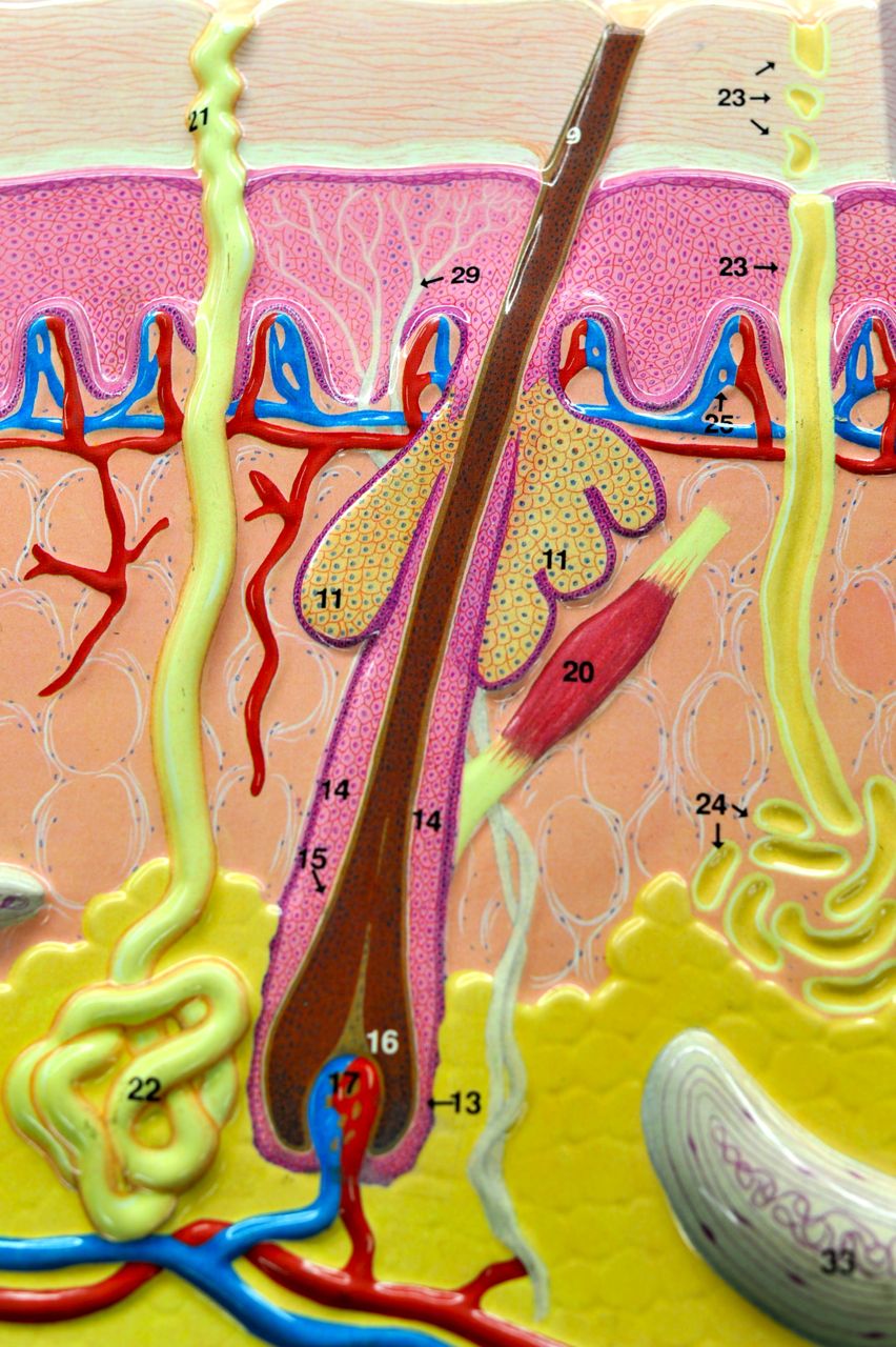
Now let's take a closer look at the structure of a hair. On the model below identify:
 Now look for the same three structures (hair bulb, arrector pili muscle, sebaceous gland) on the model below, and also try to locate:
 Hair papilla (connective tissue papilla) can only be seen in the model below. Can you see it?
In addition, the picture below shows two out of three common skin glands:
(Side note: There are also apocrine sweat glands that are present mostly in the skin of the axillary and anogenital areas. Contrary to the eccrine sweat glands shown in the picture below, those glands empty into hair follicles. They are not shown on our models.)


Let's zoom in on different parts of the model and try to figure out what all the numbers stand for.
First, let's look at skin layers. The skin has three principal layers:
- Epidermis (from Greek, "epi" means above);
- Dermis;
- Hypodermis (from Greek, "hypo" means below or low).
What does it mean that the epidermis is "nonvascular?" Can you recognize the components of the vasculature–the veins and arteries–on the model? (Side note: Arteries carry oxygenated blood to tissues, while veins drain deoxygenated blood from tissues. On all our models vessels carrying oxygenated blood are painted red, and vessels carrying oxygen-poor blood are painted blue.)
Epidermis, the outermost (or the most superficial) layer of the skin, is comprised mostly of specialized cells called keratinocytes. It can be further subdivided into four (five in the thick skin of our palms and soles) layers called strata:
- Stratum corneum
- Stratum lucidum (only present in the thick skin of our palms and soles)
- Stratum granulosum
- Stratum spinosum
- Stratum basale/germinativum
Can you identify all those layers on the model? (Clue: Focus on numbers 1 through 5.) You should also know the key differences between those skin layers.
Dermis, the middle layer of the skin, can be divided into two parts:
- Papillary layer
- Reticular layer
Let's now look at the various sensory neurons present in our skin. Can you see them on the model?
All neurons are shown in white (numbers 28 to 33). As you can see, those neurons can be present in various skin layers, and depending on the location (and type) they have slightly different functions. Some neurons are primarily involved in perception of touch, while others in perception of pain, temperature, pressure, texture, and/or stretch. You don't need to know the names of various sensory neurons, just that there are several types, and that they serve diverse functions. (If you're curious, you can read more about different sensory neurons by following these links: free nerve endings, Meissners's corpuscle, Pacinian corpuscle, Merkel nerve endings, Ruffini corpuscle.)

Now let's take a closer look at the structure of a hair. On the model below identify:
- hair bulb
- arrector pili muscle ("hair-raising muscle")
- sebaceous (oil) gland associated with the hair follicle

- hair root
- hair shaft
- hair follicle

In addition, the picture below shows two out of three common skin glands:
- sebaceous gland associated with the hair follicle
- sudoriferous (sweat) eccrine gland to the left and right of the hair follicle
(Side note: There are also apocrine sweat glands that are present mostly in the skin of the axillary and anogenital areas. Contrary to the eccrine sweat glands shown in the picture below, those glands empty into hair follicles. They are not shown on our models.)
No comments:
Post a Comment